

Osteochondrosis dissecans
Classification of Osteochondrosis dissecans
Osteochondrosis dissecans is classified according to the stage and stability of the affected fragment:
- Stage I – Early stage: In the early stage, there is reduced blood flow to the subchondral bone without detachment occurring.
- Stage II – Partial detachment: The cartilage-bone fragment begins to partially detach but remains attached to the surrounding tissue.
- Stage III – Complete detachment: The fragment is completely detached but remains in its original position in the joint.
- Stage IV – Free joint bodies: The fragment is free in the joint and can lead to blockages and mechanical discomfort.
Causes of osteochondrosis dissecans
The exact causes of osteochondrosis dissecans are not fully understood. However, it is believed that several factors play a role:
- Mechanical overload:
- Repeated microtraumas, such as those from sports activities like jumping or running, can damage the subchondral bone structure.
- Circulatory disorders:
- Local reduced blood flow to the subchondral bone leads to necrosis and promotes the detachment of the cartilage-bone fragment.
- Genetic predisposition:
- A familial clustering suggests genetic factors that increase the risk of the disease.
- Growth disorders:
- In adolescents during the growth phase, developmental disorders in the bone can promote the development of OD.
- Misalignments:
- Misloading due to joint misalignments such as knock-knees or bowlegs favors the disease.
Symptoms of osteochondrosis dissecans
The symptoms of osteochondrosis dissecans depend on the affected joint and the stage of the disease:
- Load-dependent pain: Pain initially occurs only under load but can also persist at rest in later stages.
- Swelling: Recurring swelling is a common sign, especially after physical activity.
- Movement restrictions: Limitations in joint mobility due to pain or mechanical blockages.
- Instability: A feeling of insecurity in the joint, especially in advanced stages with loose fragments.
- Crunching or rubbing: Mechanical noises that can arise from the movement of free joint bodies.
Diagnosis of osteochondrosis dissecans
The diagnosis is made through a combination of medical history, clinical examination, and imaging techniques:
- Medical history: The doctor inquires about the patient's medical history, symptoms, stress patterns, and possible injuries.
- Clinical examination: Checking for swelling, tenderness, and movement restrictions of the affected joint.
- Imaging techniques:
- X-ray: Shows bony changes and the condition of the fragment.
- MRI: Provides detailed information about the condition of the cartilage and subchondral bone.
- CT: Precise imaging of bone structures and fragments.
Treatment options for osteochondrosis dissecans
Conservative Therapy
In the early stages and in young patients, OD can often be treated without surgery:
- Relief:
- Avoidance of strenuous activities through immobilization or crutches.
- Physiotherapy:
- Exercises to strengthen muscles and improve joint stability.
- Medication Therapy:
- Use of painkillers and anti-inflammatory drugs to alleviate symptoms.
Surgical Therapy
In advanced stages or with fragment instability, surgery is often necessary:
- Drilling:
- Promoting blood circulation in the subchondral bone through small drill holes.
- Fragment Fixation:
- Reattachment of the detached fragment with screws or pins.
- Removal of Loose Joint Bodies:
- Surgical removal of detached fragments to eliminate blockages.
- Cartilage Transplantation:
- Replacement of damaged cartilage tissue with the body's own or artificial material.
Preventive measures for osteochondrosis dissecans
The following measures can help prevent the disease:
- Avoidance of overexertion: Limiting intense sports activities and taking regular breaks.
- Strengthening the muscles: Regular training to support joint stability.
- Early detection: Timely examination for recurring joint pain or swelling.
- Ergonomic stress: Optimization of movement sequences in sports and everyday life.
- Control of misalignments: Treatment of joint misalignments with insoles or physiotherapy.

Knee joint inflammation (arthritis)
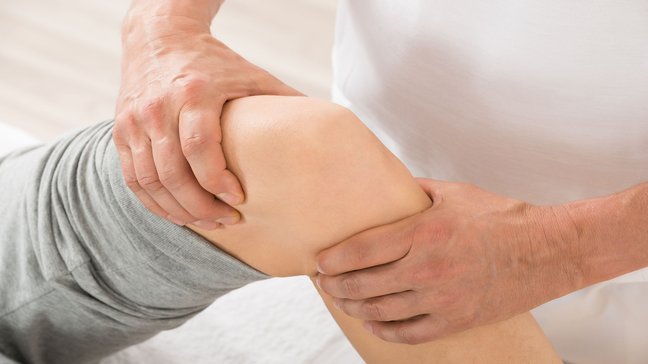
Inflammation of the knee joint, medically referred to as arthritis of the knee joint, is an inflammatory condition that affects the knee joint. It can be triggered by infections, autoimmune diseases, or overuse and leads to pain, swelling, and…
Bursitis in the knee (Bursitis)
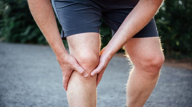
Bursitis in the knee, also known as bursitis, is an inflammatory condition of the bursae in the knee joint. These fluid-filled cushions reduce friction between bones, tendons, and muscles. There are several bursae in the knee, the most commonly…
Patellar tip syndrome (Jumper's Knee)
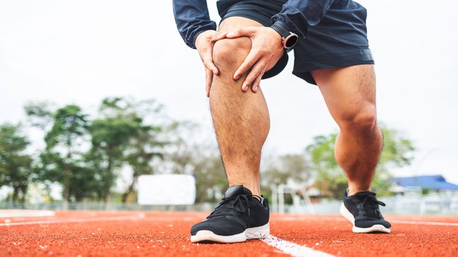
A patellar dislocation refers to the dislocation of the kneecap (patella) from its natural position in the femoral groove of the thigh bone (femur). This injury often occurs in young, active individuals and is usually associated with severe pain and…
Osgood-Schlatter disease
Osgood-Schlatter disease is a common condition in adolescents caused by overuse of the shinbone at the site of the patellar tendon attachment. It is classified as a growth disorder (osteochondrosis) and primarily affects physically active children…
Osteochondrosis dissecans
Classification of Osteochondrosis dissecans
Osteochondrosis dissecans is classified according to the stage and stability of the affected fragment:
- Stage I – Early stage: In the early stage, there is reduced blood flow to the subchondral bone without detachment occurring.
- Stage II – Partial detachment: The cartilage-bone fragment begins to partially detach but remains attached to the surrounding tissue.
- Stage III – Complete detachment: The fragment is completely detached but remains in its original position in the joint.
- Stage IV – Free joint bodies: The fragment is free in the joint and can lead to blockages and mechanical discomfort.
Causes of osteochondrosis dissecans
The exact causes of osteochondrosis dissecans are not fully understood. However, it is believed that several factors play a role:
- Mechanical overload:
- Repeated microtraumas, such as those from sports activities like jumping or running, can damage the subchondral bone structure.
- Circulatory disorders:
- Local reduced blood flow to the subchondral bone leads to necrosis and promotes the detachment of the cartilage-bone fragment.
- Genetic predisposition:
- A familial clustering suggests genetic factors that increase the risk of the disease.
- Growth disorders:
- In adolescents during the growth phase, developmental disorders in the bone can promote the development of OD.
- Misalignments:
- Misloading due to joint misalignments such as knock-knees or bowlegs favors the disease.
Symptoms of osteochondrosis dissecans
The symptoms of osteochondrosis dissecans depend on the affected joint and the stage of the disease:
- Load-dependent pain: Pain initially occurs only under load but can also persist at rest in later stages.
- Swelling: Recurring swelling is a common sign, especially after physical activity.
- Movement restrictions: Limitations in joint mobility due to pain or mechanical blockages.
- Instability: A feeling of insecurity in the joint, especially in advanced stages with loose fragments.
- Crunching or rubbing: Mechanical noises that can arise from the movement of free joint bodies.
Diagnosis of osteochondrosis dissecans
The diagnosis is made through a combination of medical history, clinical examination, and imaging techniques:
- Medical history: The doctor inquires about the patient's medical history, symptoms, stress patterns, and possible injuries.
- Clinical examination: Checking for swelling, tenderness, and movement restrictions of the affected joint.
- Imaging techniques:
- X-ray: Shows bony changes and the condition of the fragment.
- MRI: Provides detailed information about the condition of the cartilage and subchondral bone.
- CT: Precise imaging of bone structures and fragments.
Treatment options for osteochondrosis dissecans
Conservative Therapy
In the early stages and in young patients, OD can often be treated without surgery:
- Relief:
- Avoidance of strenuous activities through immobilization or crutches.
- Physiotherapy:
- Exercises to strengthen muscles and improve joint stability.
- Medication Therapy:
- Use of painkillers and anti-inflammatory drugs to alleviate symptoms.
Surgical Therapy
In advanced stages or with fragment instability, surgery is often necessary:
- Drilling:
- Promoting blood circulation in the subchondral bone through small drill holes.
- Fragment Fixation:
- Reattachment of the detached fragment with screws or pins.
- Removal of Loose Joint Bodies:
- Surgical removal of detached fragments to eliminate blockages.
- Cartilage Transplantation:
- Replacement of damaged cartilage tissue with the body's own or artificial material.
Preventive measures for osteochondrosis dissecans
The following measures can help prevent the disease:
- Avoidance of overexertion: Limiting intense sports activities and taking regular breaks.
- Strengthening the muscles: Regular training to support joint stability.
- Early detection: Timely examination for recurring joint pain or swelling.
- Ergonomic stress: Optimization of movement sequences in sports and everyday life.
- Control of misalignments: Treatment of joint misalignments with insoles or physiotherapy.
Knee joint inflammation (arthritis)
Inflammation of the knee joint, medically referred to as arthritis of the knee joint, is an inflammatory condition that affects the knee joint. It can be triggered by infections, autoimmune diseases, or overuse and leads to pain, swelling, and…
Bursitis in the knee (Bursitis)
Bursitis in the knee, also known as bursitis, is an inflammatory condition of the bursae in the knee joint. These fluid-filled cushions reduce friction between bones, tendons, and muscles. There are several bursae in the knee, the most commonly…
Patellar tip syndrome (Jumper's Knee)
A patellar dislocation refers to the dislocation of the kneecap (patella) from its natural position in the femoral groove of the thigh bone (femur). This injury often occurs in young, active individuals and is usually associated with severe pain and…
Osgood-Schlatter disease
Osgood-Schlatter disease is a common condition in adolescents caused by overuse of the shinbone at the site of the patellar tendon attachment. It is classified as a growth disorder (osteochondrosis) and primarily affects physically active children…