

Patellar luxation
Classification of Patellar Luxations
Patellar dislocations are categorized according to cause and progression as follows:
- Acute traumatic dislocation: Occurs as a result of direct trauma or sudden force impact.
- Recurrent dislocation: Repeated dislocation of the kneecap, often due to misalignments or unstable ligaments.
- Congenital dislocation: Congenital misalignment of the kneecap, diagnosed in childhood.
Causes of patellar luxation
The causes of a patellar dislocation are varied and can be mechanical, anatomical, or genetic in nature:
- Traumatic events:
- Sudden lateral force, such as from a fall or blow to the knee.
- Anatomical factors:
- Misalignments such as knock-knees, a high patella position (patella alta), or a shallow trochlea (patellar groove).
- Ligament instability:
- Weakness or injuries of the medial patellofemoral ligament (MPFL), which stabilizes the patella.
- Muscle imbalances:
- Uneven pulling forces of the thigh muscles, which can pull the patella out of position.
- Genetic predisposition:
- Familial occurrence of patellar dislocations suggests a genetic component.
Symptoms of patellar luxation
The symptoms of a patellar dislocation are characteristic and include:
- Acute pain: Severe pain in the area of the kneecap, especially immediately after the dislocation.
- Deformity: Visible displacement of the patella, usually to the outside of the knee.
- Swelling: Rapid swelling due to bruising in the joint (hemarthrosis).
- Instability: Feeling of "giving way" or insecurity in the knee, especially during movement.
- Limited range of motion: Difficulty in extending or bending the knee.
Diagnosis of patellar dislocation
The diagnosis is made through medical history, clinical examination, and imaging techniques:
1. **Medical History:** Collection of the patient's medical history, description of symptoms, and circumstances of the injury.
2. **Clinical Examination:**
- Checking the position of the kneecap and the stability of the surrounding structures.
- Tests such as the apprehension test, where lateral displacement pressure on the patella is checked.
3. **Imaging Techniques:**
- **X-ray:** Visualization of bone injuries and misalignments.
- **MRI:** Examination of soft tissue damage, such as injuries to the MPFL or cartilage damage.
- **CT:** Detailed assessment of anatomical deviations and fractures.
Treatment options for patellar luxation
Conservative Therapy
In cases of initial or mild patellar dislocation, conservative treatment may be sufficient:
- Reposition:
- Manual repositioning of the patella to its natural position, usually by a doctor.
- Immobilization:
- Use of knee braces or splints to stabilize the joint and promote healing.
- Physiotherapy:
- Exercises to strengthen the thigh muscles and improve stability.
- Cold Applications:
- Reduction of pain and swelling through cold packs.
- Medication Therapy:
- Intake of NSAIDs for pain relief and anti-inflammation.
Surgical Therapy
In cases of repeated dislocations or significant damage, surgical intervention may be necessary:
- MPFL Reconstruction:
- Restoration or reinforcement of the medial patellofemoral ligament.
- Trochleoplasty:
- Surgical deepening of the groove to improve patellar tracking.
- Tibial Tuberosity Transfer:
- Modification of the patellar tendon attachment site to correct the pull direction.
- Cartilage Reconstruction:
- Treatment of accompanying injuries such as cartilage damage.
Preventive measures for patellar dislocation
The following measures are helpful in preventing a patellar dislocation:
- Strengthening the muscles: Regular training of the thigh muscles to stabilize the kneecap.
- Avoiding overexertion: Limiting intense sports activities, especially those with sudden changes in direction.
- Correction of misalignments: Use of insoles or orthoses to improve leg alignment.
- Coordination training: Exercises to improve balance and proprioception.
- Early detection and treatment: Timely examination at the first signs of instability or pain in the knee.
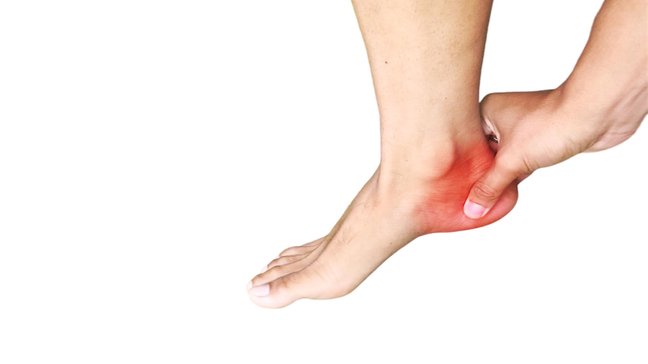
Calcaneal apophysitis
Apophysitis calcanei, also known as Sever’s disease, is an inflammatory condition of the growth plate (apophysis) of the heel bone. It occurs predominantly in children and adolescents aged 8 to 16 years, especially during growth phases. Those…
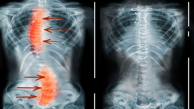
Scoliosis
Scoliosis refers to a lateral curvature of the spine that is accompanied by a rotation of the vertebral bodies. This misalignment can occur in any section of the spine, but most commonly in the thoracic and lumbar spine. Scoliosis occurs in both…

Scheuermann's disease
Morbus Scheuermann, auch als Scheuermann-Krankheit bezeichnet, ist eine Wachstumsstörung der Wirbelsäule, die vor allem bei Jugendlichen während der Wachstumsphase auftritt. Die Erkrankung führt zu einer vermehrten Krümmung der Brustwirbelsäule…
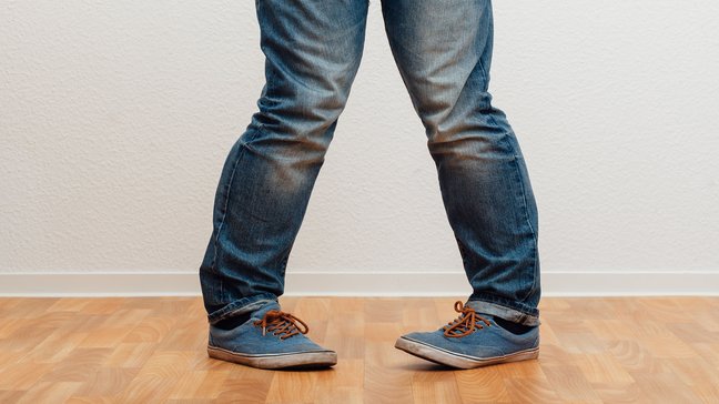
Misalignments of the leg axes
Misalignments of the leg axes are deviations from the normal alignment of the lower limbs. They can significantly affect the way weight is distributed across the joints, potentially leading to pain, reduced mobility, and long-term damage,…
Patellar luxation
Classification of Patellar Luxations
Patellar dislocations are categorized according to cause and progression as follows:
- Acute traumatic dislocation: Occurs as a result of direct trauma or sudden force impact.
- Recurrent dislocation: Repeated dislocation of the kneecap, often due to misalignments or unstable ligaments.
- Congenital dislocation: Congenital misalignment of the kneecap, diagnosed in childhood.
Causes of patellar luxation
The causes of a patellar dislocation are varied and can be mechanical, anatomical, or genetic in nature:
- Traumatic events:
- Sudden lateral force, such as from a fall or blow to the knee.
- Anatomical factors:
- Misalignments such as knock-knees, a high patella position (patella alta), or a shallow trochlea (patellar groove).
- Ligament instability:
- Weakness or injuries of the medial patellofemoral ligament (MPFL), which stabilizes the patella.
- Muscle imbalances:
- Uneven pulling forces of the thigh muscles, which can pull the patella out of position.
- Genetic predisposition:
- Familial occurrence of patellar dislocations suggests a genetic component.
Symptoms of patellar luxation
The symptoms of a patellar dislocation are characteristic and include:
- Acute pain: Severe pain in the area of the kneecap, especially immediately after the dislocation.
- Deformity: Visible displacement of the patella, usually to the outside of the knee.
- Swelling: Rapid swelling due to bruising in the joint (hemarthrosis).
- Instability: Feeling of "giving way" or insecurity in the knee, especially during movement.
- Limited range of motion: Difficulty in extending or bending the knee.
Diagnosis of patellar dislocation
The diagnosis is made through medical history, clinical examination, and imaging techniques:
1. **Medical History:** Collection of the patient's medical history, description of symptoms, and circumstances of the injury.
2. **Clinical Examination:**
- Checking the position of the kneecap and the stability of the surrounding structures.
- Tests such as the apprehension test, where lateral displacement pressure on the patella is checked.
3. **Imaging Techniques:**
- **X-ray:** Visualization of bone injuries and misalignments.
- **MRI:** Examination of soft tissue damage, such as injuries to the MPFL or cartilage damage.
- **CT:** Detailed assessment of anatomical deviations and fractures.
Treatment options for patellar luxation
Conservative Therapy
In cases of initial or mild patellar dislocation, conservative treatment may be sufficient:
- Reposition:
- Manual repositioning of the patella to its natural position, usually by a doctor.
- Immobilization:
- Use of knee braces or splints to stabilize the joint and promote healing.
- Physiotherapy:
- Exercises to strengthen the thigh muscles and improve stability.
- Cold Applications:
- Reduction of pain and swelling through cold packs.
- Medication Therapy:
- Intake of NSAIDs for pain relief and anti-inflammation.
Surgical Therapy
In cases of repeated dislocations or significant damage, surgical intervention may be necessary:
- MPFL Reconstruction:
- Restoration or reinforcement of the medial patellofemoral ligament.
- Trochleoplasty:
- Surgical deepening of the groove to improve patellar tracking.
- Tibial Tuberosity Transfer:
- Modification of the patellar tendon attachment site to correct the pull direction.
- Cartilage Reconstruction:
- Treatment of accompanying injuries such as cartilage damage.
Preventive measures for patellar dislocation
The following measures are helpful in preventing a patellar dislocation:
- Strengthening the muscles: Regular training of the thigh muscles to stabilize the kneecap.
- Avoiding overexertion: Limiting intense sports activities, especially those with sudden changes in direction.
- Correction of misalignments: Use of insoles or orthoses to improve leg alignment.
- Coordination training: Exercises to improve balance and proprioception.
- Early detection and treatment: Timely examination at the first signs of instability or pain in the knee.
Calcaneal apophysitis
Apophysitis calcanei, also known as Sever’s disease, is an inflammatory condition of the growth plate (apophysis) of the heel bone. It occurs predominantly in children and adolescents aged 8 to 16 years, especially during growth phases. Those…
Scoliosis
Scoliosis refers to a lateral curvature of the spine that is accompanied by a rotation of the vertebral bodies. This misalignment can occur in any section of the spine, but most commonly in the thoracic and lumbar spine. Scoliosis occurs in both…
Scheuermann's disease
Morbus Scheuermann, auch als Scheuermann-Krankheit bezeichnet, ist eine Wachstumsstörung der Wirbelsäule, die vor allem bei Jugendlichen während der Wachstumsphase auftritt. Die Erkrankung führt zu einer vermehrten Krümmung der Brustwirbelsäule…
Misalignments of the leg axes
Misalignments of the leg axes are deviations from the normal alignment of the lower limbs. They can significantly affect the way weight is distributed across the joints, potentially leading to pain, reduced mobility, and long-term damage,…