

Upper heel spur
Classification of upper heel spur
Upper heel spurs can be classified depending on the cause and manifestation:
- Mechanical heel spur: Caused by chronic friction or pressure, such as from tight shoes.
- Structural heel spur: Changes in bone shape due to congenital or growth disorders.
- Inflammatory heel spur: Accompanying inflammatory processes such as Achilles tendinitis.
Causes of upper heel spur
The main causes of a superior heel spur are:
- Mechanical overload:
- Persistent pressure or friction from unsuitable footwear.
- Misalignments:
- Foot misalignments such as a high arch or flat foot increase pressure on the heel region.
- Excessive athletic activity:
- Intensive strains, especially from running or jumping, can promote development.
- Genetic factors:
- A familial predisposition can influence bone structure and thus the formation of a superior heel spur.
Symptoms of upper heel spur
The symptoms of an upper heel spur are characteristic and include:
- Local pain: Pain at the upper back of the heel, especially with pressure or movement.
- Swelling: Visible and palpable swelling in the area of the Achilles tendon.
- Redness and irritation: Skin irritations due to friction, often caused by shoes.
- Movement restrictions: Pain when walking, especially during sports activities or barefoot on hard floors.
- Pressure sensitivity: Increased pain when touching or pressing on the affected area.
Diagnosis of upper heel spur
The diagnosis is based on a combination of medical history, clinical examination, and imaging techniques:
- Medical History:
- The doctor inquires about typical symptoms, possible triggers, and shoe habits.
- Clinical Examination:
- Visual and tactile findings to identify the bony protrusion and painful areas.
- Imaging Techniques:
- X-ray: Visualization of bony changes and size of the heel spur.
- MRI: Detailed assessment of soft tissue damage and inflammation.
- Ultrasound: Examination of the Achilles tendon and adjacent structures.
Treatment options for upper heel spur
Conservative Therapy
Most cases can be treated without surgery:
- Shoe Adjustment:
- Avoiding tight, pressing shoes and using shoes with soft padding in the heel area.
- Orthopedic Insoles:
- Special insoles for pressure relief and improvement of foot positioning.
- Physiotherapy:
- Exercises to strengthen the calf muscles and relieve the Achilles tendon.
- Cold or Heat Applications:
- Relief of pain and inflammation through targeted applications.
- Medication Therapy:
- Taking NSAIDs to reduce pain and inflammation.
Surgical Therapy
If conservative measures are not sufficient, surgery may be necessary:
- Removal of Heel Spur:
- Removal of the bony protrusion for pressure relief.
- Achilles Tendon Correction:
- If the tendon is also involved, it can be treated surgically.
Preventive measures for upper heel spur
To prevent a superior heel spur, the following measures are helpful:
- Suitable footwear: Wearing shoes with sufficient space and soft padding in the heel area.
- Avoiding overstrain: Limiting intense sports activities and regular recovery phases.
- Strengthening the muscles: Specific exercises to strengthen the calf and foot muscles.
- Regular exercise: Promoting blood circulation and relieving the heel area through joint-friendly sports such as swimming or cycling.
- Early detection: Timely response to the first signs of pain or swelling.
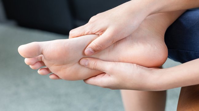
Metatarsalgia
Metatarsalgia describes pain in the forefoot area, often occurring under the metatarsal heads. These complaints usually arise from overuse, misalignments, or degenerative changes in the foot. People who stand a lot, walk, or are physically active are…
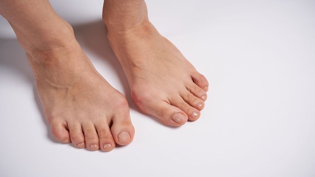
Hallux rigidus
Hallux rigidus is osteoarthritis of the big toe joint, which can lead to painful movement restrictions and eventually to joint stiffening. This condition significantly impairs the rolling motion of the foot and often affects people from middle age…
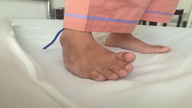
Clubfoot (Pes Equinovarus)
Clubfoot (Pes equinovarus) is a complex congenital foot deformity that occurs in about 1 in 1,000 newborns worldwide. Boys are more frequently affected than girls, and in about 50% of cases, the clubfoot is bilateral. This deformity, which can lead…
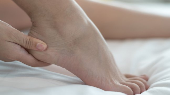
Tarsal tunnel syndrome
Tarsal tunnel syndrome is a nerve compression disorder in which the tibial nerve is constricted in the area of the tarsal tunnel, an anatomical structure on the inside of the foot. This compression leads to pain, sensory disturbances, and possibly…
Upper heel spur
Classification of upper heel spur
Upper heel spurs can be classified depending on the cause and manifestation:
- Mechanical heel spur: Caused by chronic friction or pressure, such as from tight shoes.
- Structural heel spur: Changes in bone shape due to congenital or growth disorders.
- Inflammatory heel spur: Accompanying inflammatory processes such as Achilles tendinitis.
Causes of upper heel spur
The main causes of a superior heel spur are:
- Mechanical overload:
- Persistent pressure or friction from unsuitable footwear.
- Misalignments:
- Foot misalignments such as a high arch or flat foot increase pressure on the heel region.
- Excessive athletic activity:
- Intensive strains, especially from running or jumping, can promote development.
- Genetic factors:
- A familial predisposition can influence bone structure and thus the formation of a superior heel spur.
Symptoms of upper heel spur
The symptoms of an upper heel spur are characteristic and include:
- Local pain: Pain at the upper back of the heel, especially with pressure or movement.
- Swelling: Visible and palpable swelling in the area of the Achilles tendon.
- Redness and irritation: Skin irritations due to friction, often caused by shoes.
- Movement restrictions: Pain when walking, especially during sports activities or barefoot on hard floors.
- Pressure sensitivity: Increased pain when touching or pressing on the affected area.
Diagnosis of upper heel spur
The diagnosis is based on a combination of medical history, clinical examination, and imaging techniques:
- Medical History:
- The doctor inquires about typical symptoms, possible triggers, and shoe habits.
- Clinical Examination:
- Visual and tactile findings to identify the bony protrusion and painful areas.
- Imaging Techniques:
- X-ray: Visualization of bony changes and size of the heel spur.
- MRI: Detailed assessment of soft tissue damage and inflammation.
- Ultrasound: Examination of the Achilles tendon and adjacent structures.
Treatment options for upper heel spur
Conservative Therapy
Most cases can be treated without surgery:
- Shoe Adjustment:
- Avoiding tight, pressing shoes and using shoes with soft padding in the heel area.
- Orthopedic Insoles:
- Special insoles for pressure relief and improvement of foot positioning.
- Physiotherapy:
- Exercises to strengthen the calf muscles and relieve the Achilles tendon.
- Cold or Heat Applications:
- Relief of pain and inflammation through targeted applications.
- Medication Therapy:
- Taking NSAIDs to reduce pain and inflammation.
Surgical Therapy
If conservative measures are not sufficient, surgery may be necessary:
- Removal of Heel Spur:
- Removal of the bony protrusion for pressure relief.
- Achilles Tendon Correction:
- If the tendon is also involved, it can be treated surgically.
Preventive measures for upper heel spur
To prevent a superior heel spur, the following measures are helpful:
- Suitable footwear: Wearing shoes with sufficient space and soft padding in the heel area.
- Avoiding overstrain: Limiting intense sports activities and regular recovery phases.
- Strengthening the muscles: Specific exercises to strengthen the calf and foot muscles.
- Regular exercise: Promoting blood circulation and relieving the heel area through joint-friendly sports such as swimming or cycling.
- Early detection: Timely response to the first signs of pain or swelling.
Metatarsalgia
Metatarsalgia describes pain in the forefoot area, often occurring under the metatarsal heads. These complaints usually arise from overuse, misalignments, or degenerative changes in the foot. People who stand a lot, walk, or are physically active are…
Hallux rigidus
Hallux rigidus is osteoarthritis of the big toe joint, which can lead to painful movement restrictions and eventually to joint stiffening. This condition significantly impairs the rolling motion of the foot and often affects people from middle age…
Clubfoot (Pes Equinovarus)
Clubfoot (Pes equinovarus) is a complex congenital foot deformity that occurs in about 1 in 1,000 newborns worldwide. Boys are more frequently affected than girls, and in about 50% of cases, the clubfoot is bilateral. This deformity, which can lead…
Tarsal tunnel syndrome
Tarsal tunnel syndrome is a nerve compression disorder in which the tibial nerve is constricted in the area of the tarsal tunnel, an anatomical structure on the inside of the foot. This compression leads to pain, sensory disturbances, and possibly…