

Osteonecrosis in childhood
Classification of Osteonecrosis in Childhood
Osteonecroses in childhood can be classified according to location and cause:
1. **Legg-Calvé-Perthes disease:** Necrosis of the femoral head, common in children between 4 and 10 years old.
2. **Osgood-Schlatter disease:** Necrosis of the tibial tuberosity, typical in physically active adolescents.
3. **Kienböck's disease:** Necrosis of the lunate bone in the wrist.
4. **Köhler disease I:** Necrosis of the navicular bone in the foot.
5. **Panner's disease:** Necrosis of the humeral head, especially in young athletes.
Causes of Osteonecrosis in Childhood
The exact causes are often not fully clarified, but various factors can contribute to the development:
- Circulatory disorders:
- Reduced blood supply to the growth zone leads to tissue loss.
- Mechanical overload:
- Excessive strain during sports activities can damage the vessels in the growth zone.
- Traumatic events:
- Injuries such as falls or fractures can cause circulatory disorders.
- Systemic diseases:
- Disorders such as sickle cell anemia or coagulation disorders increase the risk of osteonecrosis.
- Genetic predisposition:
- Familial predisposition can increase the likelihood.
Symptoms of osteonecrosis in childhood
The symptoms vary depending on the affected region and stage of the disease:
- **Pain:** Load-dependent pain in the affected area, which may decrease at rest.
- **Swelling:** Local swelling, especially in the lower extremities.
- **Movement restrictions:** Reduced mobility of the affected joint, often gradually.
- **Gait changes:** Limping or protective postures in osteonecrosis of the lower extremities.
- **Muscle atrophy:** Decrease in muscle mass due to protection and reduced activity.
Diagnosis of osteonecrosis in childhood
The diagnosis is based on a combination of medical history, clinical examination, and imaging techniques:
- Medical History: The doctor inquires about symptoms, previous illnesses, and possible injuries.
- Clinical Examination: Checking for tenderness, swelling, and movement restrictions in the affected area.
- Imaging Techniques:
- X-ray: Early stages often unremarkable, later changes in bone structure visible.
- MRI: Highly sensitive method for detecting circulatory disorders.
- CT: Detailed depiction of bone changes.
Treatment options for osteonecrosis in childhood
Conservative Therapy
In most cases, treatment can be done without surgery, especially in the early stages:
- Relief:
- Avoidance of stressful activities through immobilization or crutches.
- Physiotherapy:
- Exercises to strengthen muscles and promote mobility.
- Pain Therapy:
- Use of NSAIDs to relieve pain and inflammation.
- Orthopedic Aids:
- Insoles or orthoses for stabilization and pressure distribution.
Surgical Therapy
In advanced stages or persistent complaints, surgical intervention may be necessary:
- Corrective Osteotomy:
- Change of bone axis to relieve the affected area.
- Drilling:
- Promotion of blood circulation through small drill holes in the bone.
- Transplantation:
- Replacement of necrotic tissue with bone or cartilage grafts.
Preventive measures for osteonecrosis in childhood
To prevent osteonecrosis in childhood, the following measures can contribute:
- Avoidance of overstrain: Regular breaks and appropriate strain during sports.
- Early treatment of injuries: Timely care for trauma or swelling.
- Regular exercise: Promotion of blood circulation and strengthening of muscles through gentle activities.
- Nutritional optimization: Ensuring adequate supply of calcium and vitamin D.
- Medical check-ups: Regular examinations for pre-existing conditions that may increase the risk.
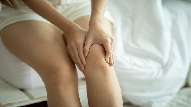
Patellar luxation
Eine Patellaluxation bezeichnet das Herausspringen der Kniescheibe (Patella) aus ihrer natürlichen Position im Gleitlager des Oberschenkelknochens (Femur). Diese Verletzung tritt häufig bei jungen, aktiven Menschen auf und ist meist mit starken…
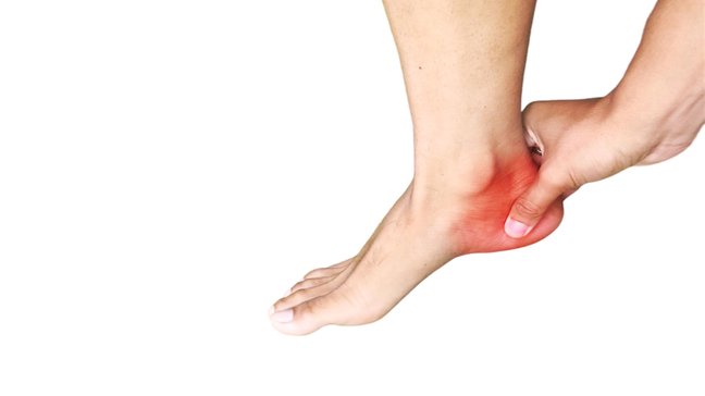
Calcaneal apophysitis
Apophysitis calcanei, also known as Sever’s disease, is an inflammatory condition of the growth plate (apophysis) of the heel bone. It occurs predominantly in children and adolescents aged 8 to 16 years, especially during growth phases. Those…
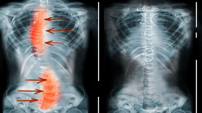
Scoliosis
Scoliosis refers to a lateral curvature of the spine that is accompanied by a rotation of the vertebral bodies. This misalignment can occur in any section of the spine, but most commonly in the thoracic and lumbar spine. Scoliosis occurs in both…
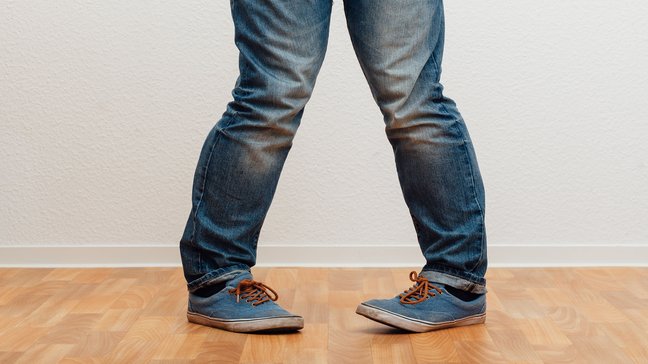
Misalignments of the leg axes
Misalignments of the leg axes are deviations from the normal alignment of the lower limbs. They can significantly affect the way weight is distributed across the joints, potentially leading to pain, reduced mobility, and long-term damage,…
Osteonecrosis in childhood
Classification of Osteonecrosis in Childhood
Osteonecroses in childhood can be classified according to location and cause:
1. **Legg-Calvé-Perthes disease:** Necrosis of the femoral head, common in children between 4 and 10 years old.
2. **Osgood-Schlatter disease:** Necrosis of the tibial tuberosity, typical in physically active adolescents.
3. **Kienböck's disease:** Necrosis of the lunate bone in the wrist.
4. **Köhler disease I:** Necrosis of the navicular bone in the foot.
5. **Panner's disease:** Necrosis of the humeral head, especially in young athletes.
Causes of Osteonecrosis in Childhood
The exact causes are often not fully clarified, but various factors can contribute to the development:
- Circulatory disorders:
- Reduced blood supply to the growth zone leads to tissue loss.
- Mechanical overload:
- Excessive strain during sports activities can damage the vessels in the growth zone.
- Traumatic events:
- Injuries such as falls or fractures can cause circulatory disorders.
- Systemic diseases:
- Disorders such as sickle cell anemia or coagulation disorders increase the risk of osteonecrosis.
- Genetic predisposition:
- Familial predisposition can increase the likelihood.
Symptoms of osteonecrosis in childhood
The symptoms vary depending on the affected region and stage of the disease:
- **Pain:** Load-dependent pain in the affected area, which may decrease at rest.
- **Swelling:** Local swelling, especially in the lower extremities.
- **Movement restrictions:** Reduced mobility of the affected joint, often gradually.
- **Gait changes:** Limping or protective postures in osteonecrosis of the lower extremities.
- **Muscle atrophy:** Decrease in muscle mass due to protection and reduced activity.
Diagnosis of osteonecrosis in childhood
The diagnosis is based on a combination of medical history, clinical examination, and imaging techniques:
- Medical History: The doctor inquires about symptoms, previous illnesses, and possible injuries.
- Clinical Examination: Checking for tenderness, swelling, and movement restrictions in the affected area.
- Imaging Techniques:
- X-ray: Early stages often unremarkable, later changes in bone structure visible.
- MRI: Highly sensitive method for detecting circulatory disorders.
- CT: Detailed depiction of bone changes.
Treatment options for osteonecrosis in childhood
Conservative Therapy
In most cases, treatment can be done without surgery, especially in the early stages:
- Relief:
- Avoidance of stressful activities through immobilization or crutches.
- Physiotherapy:
- Exercises to strengthen muscles and promote mobility.
- Pain Therapy:
- Use of NSAIDs to relieve pain and inflammation.
- Orthopedic Aids:
- Insoles or orthoses for stabilization and pressure distribution.
Surgical Therapy
In advanced stages or persistent complaints, surgical intervention may be necessary:
- Corrective Osteotomy:
- Change of bone axis to relieve the affected area.
- Drilling:
- Promotion of blood circulation through small drill holes in the bone.
- Transplantation:
- Replacement of necrotic tissue with bone or cartilage grafts.
Preventive measures for osteonecrosis in childhood
To prevent osteonecrosis in childhood, the following measures can contribute:
- Avoidance of overstrain: Regular breaks and appropriate strain during sports.
- Early treatment of injuries: Timely care for trauma or swelling.
- Regular exercise: Promotion of blood circulation and strengthening of muscles through gentle activities.
- Nutritional optimization: Ensuring adequate supply of calcium and vitamin D.
- Medical check-ups: Regular examinations for pre-existing conditions that may increase the risk.
Patellar luxation
Eine Patellaluxation bezeichnet das Herausspringen der Kniescheibe (Patella) aus ihrer natürlichen Position im Gleitlager des Oberschenkelknochens (Femur). Diese Verletzung tritt häufig bei jungen, aktiven Menschen auf und ist meist mit starken…
Calcaneal apophysitis
Apophysitis calcanei, also known as Sever’s disease, is an inflammatory condition of the growth plate (apophysis) of the heel bone. It occurs predominantly in children and adolescents aged 8 to 16 years, especially during growth phases. Those…
Scoliosis
Scoliosis refers to a lateral curvature of the spine that is accompanied by a rotation of the vertebral bodies. This misalignment can occur in any section of the spine, but most commonly in the thoracic and lumbar spine. Scoliosis occurs in both…
Misalignments of the leg axes
Misalignments of the leg axes are deviations from the normal alignment of the lower limbs. They can significantly affect the way weight is distributed across the joints, potentially leading to pain, reduced mobility, and long-term damage,…