

Pseudogout
Classification of Pseudogout
Pseudogout can be divided by course and cause:
- Acute pseudogout:
- Sudden, painful joint inflammations reminiscent of an acute gout attack.
- Chronic pseudogout:
- Long-lasting joint inflammations with recurring episodes that can impair joint function.
- Asymptomatic chondrocalcinosis:
- Deposits of CPP crystals without noticeable symptoms, often discovered incidentally on X-rays.
Causes of pseudogout
The main cause of pseudogout is the deposition of calcium pyrophosphate crystals in the joints. These can be promoted by various factors:
- **Age:**
- The risk of crystal formation increases with age.
- **Hereditary factors:**
- A familial predisposition can promote the development of pseudogout.
- **Underlying conditions:**
- Conditions such as hyperparathyroidism, hemochromatosis, or hypomagnesemia promote crystal formation.
- **Joint damage:**
- Pre-existing osteoarthritis or previous injuries increase the risk.
Symptoms of pseudogout
The symptoms often resemble those of gout but differ in location and progression:
- Sudden joint pain:
- Usually in a single large joint, such as the knee or wrist.
- Swelling and redness:
- The affected joint is visibly swollen, red, and overheated.
- Movement restrictions:
- Pain and swelling lead to limited mobility.
- Recurring episodes:
- The condition can recur at irregular intervals.
- Chronic complaints:
- With a prolonged course, permanent joint damage can occur.
Diagnosis of pseudogout
The diagnosis is made through medical history, clinical examination, and specific tests:
- Medical history:
- Recording of joint complaints, flare-ups, and possible pre-existing conditions.
- Physical examination:
- Checking the affected joints for swelling, redness, and movement restrictions.
- Laboratory tests:
- Inflammatory markers such as CRP and ESR are often elevated.
- Analysis of joint fluid: Detection of CPP crystals under the microscope.
- Imaging techniques:
- X-ray: Detection of calcifications in the joints.
- Ultrasound: Visualization of inflammatory processes and crystal deposits.
Treatment options for pseudogout
Conservative Therapy
The treatment focuses on alleviating symptoms and preventing further flare-ups:
- Medication Therapy:
- NSAIDs to relieve pain and inflammation.
- Corticosteroid injections for severe discomfort.
- Colchicine to prevent and treat acute flare-ups.
- Physiotherapy:
- Promoting joint mobility and preventing stiffness.
- Cold Applications:
- Cooling the affected joints for pain relief.
Surgical Therapy
Surgical interventions are rarely necessary but may be required for severe joint damage:
- Joint Lavage:
- Removal of crystal deposits from the joint.
- Endoprosthetics:
- Replacement of the joint in case of irreparable damage.
Preventive measures for pseudogout
The following measures are helpful in preventing pseudogout:
- Treatment of underlying conditions: Control of metabolic disorders such as hyperparathyroidism or hemochromatosis.
- Balanced diet: Avoidance of excess calcium or other minerals.
- Regular exercise: Promotion of joint health through joint-friendly activities such as swimming.
- Early detection: Regular medical check-ups for at-risk patients.
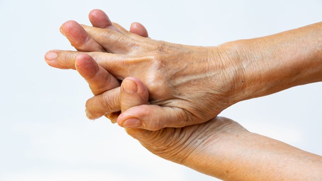
Gout
Gout is a metabolic disorder characterized by elevated uric acid levels (hyperuricemia) in the blood. It leads to the formation of uric acid crystals in joints, tissues, and organs, causing severe pain and inflammatory reactions. Gout usually occurs…
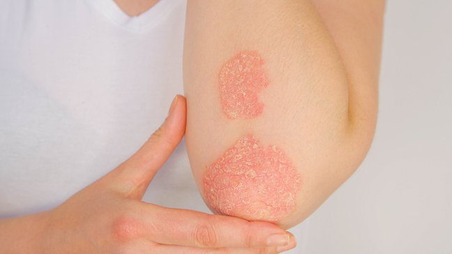
Psoriatic arthritis
Psoriatic arthritis is a chronic inflammatory disease of the joints that occurs in connection with the skin condition psoriasis. It can lead to pain, restricted movement, and permanent joint damage if not treated in a timely manner. The disease often…

Spondylarthritis
Spondyloarthritis is an inflammatory disease that can affect the spine, the sacroiliac joints, and peripheral joints. It belongs to the group of spondyloarthritides, which also includes diseases such as ankylosing spondylitis, reactive arthritis, and…

Rheumatoid arthritis
Rheumatoid arthritis (RA) is a chronic inflammatory autoimmune disease that primarily affects the joints but can also damage other organs. If left untreated, the disease leads to progressive joint destruction, pain, and movement restrictions. Women…
Pseudogout
Classification of Pseudogout
Pseudogout can be divided by course and cause:
- Acute pseudogout:
- Sudden, painful joint inflammations reminiscent of an acute gout attack.
- Chronic pseudogout:
- Long-lasting joint inflammations with recurring episodes that can impair joint function.
- Asymptomatic chondrocalcinosis:
- Deposits of CPP crystals without noticeable symptoms, often discovered incidentally on X-rays.
Causes of pseudogout
The main cause of pseudogout is the deposition of calcium pyrophosphate crystals in the joints. These can be promoted by various factors:
- **Age:**
- The risk of crystal formation increases with age.
- **Hereditary factors:**
- A familial predisposition can promote the development of pseudogout.
- **Underlying conditions:**
- Conditions such as hyperparathyroidism, hemochromatosis, or hypomagnesemia promote crystal formation.
- **Joint damage:**
- Pre-existing osteoarthritis or previous injuries increase the risk.
Symptoms of pseudogout
The symptoms often resemble those of gout but differ in location and progression:
- Sudden joint pain:
- Usually in a single large joint, such as the knee or wrist.
- Swelling and redness:
- The affected joint is visibly swollen, red, and overheated.
- Movement restrictions:
- Pain and swelling lead to limited mobility.
- Recurring episodes:
- The condition can recur at irregular intervals.
- Chronic complaints:
- With a prolonged course, permanent joint damage can occur.
Diagnosis of pseudogout
The diagnosis is made through medical history, clinical examination, and specific tests:
- Medical history:
- Recording of joint complaints, flare-ups, and possible pre-existing conditions.
- Physical examination:
- Checking the affected joints for swelling, redness, and movement restrictions.
- Laboratory tests:
- Inflammatory markers such as CRP and ESR are often elevated.
- Analysis of joint fluid: Detection of CPP crystals under the microscope.
- Imaging techniques:
- X-ray: Detection of calcifications in the joints.
- Ultrasound: Visualization of inflammatory processes and crystal deposits.
Treatment options for pseudogout
Conservative Therapy
The treatment focuses on alleviating symptoms and preventing further flare-ups:
- Medication Therapy:
- NSAIDs to relieve pain and inflammation.
- Corticosteroid injections for severe discomfort.
- Colchicine to prevent and treat acute flare-ups.
- Physiotherapy:
- Promoting joint mobility and preventing stiffness.
- Cold Applications:
- Cooling the affected joints for pain relief.
Surgical Therapy
Surgical interventions are rarely necessary but may be required for severe joint damage:
- Joint Lavage:
- Removal of crystal deposits from the joint.
- Endoprosthetics:
- Replacement of the joint in case of irreparable damage.
Preventive measures for pseudogout
The following measures are helpful in preventing pseudogout:
- Treatment of underlying conditions: Control of metabolic disorders such as hyperparathyroidism or hemochromatosis.
- Balanced diet: Avoidance of excess calcium or other minerals.
- Regular exercise: Promotion of joint health through joint-friendly activities such as swimming.
- Early detection: Regular medical check-ups for at-risk patients.
Gout
Gout is a metabolic disorder characterized by elevated uric acid levels (hyperuricemia) in the blood. It leads to the formation of uric acid crystals in joints, tissues, and organs, causing severe pain and inflammatory reactions. Gout usually occurs…
Psoriatic arthritis
Psoriatic arthritis is a chronic inflammatory disease of the joints that occurs in connection with the skin condition psoriasis. It can lead to pain, restricted movement, and permanent joint damage if not treated in a timely manner. The disease often…
Spondylarthritis
Spondyloarthritis is an inflammatory disease that can affect the spine, the sacroiliac joints, and peripheral joints. It belongs to the group of spondyloarthritides, which also includes diseases such as ankylosing spondylitis, reactive arthritis, and…
Rheumatoid arthritis
Rheumatoid arthritis (RA) is a chronic inflammatory autoimmune disease that primarily affects the joints but can also damage other organs. If left untreated, the disease leads to progressive joint destruction, pain, and movement restrictions. Women…