

Clubfoot (Pes Equinovarus)
Classification of Clubfoot
Clubfoot is divided into different categories, depending on the cause and severity:
- Idiopathic clubfoot:
- Most common form with no known cause.
- Syndromic clubfoot:
- Occurs in connection with genetic syndromes such as arthrogryposis or trisomy 18.
- Neuropathic clubfoot:
- Caused by neurological disorders such as spina bifida.
- Postural clubfoot:
- Result of fetal position anomalies in the womb.
Causes of clubfoot
The exact causes of clubfoot are not fully understood. However, various factors play a role:
- Genetic predisposition:
- Clubfoot occurs more frequently in families, indicating a genetic component.
- Mechanical factors:
- Lack of space in the womb or an unfavorable position of the fetus can affect the development of the foot.
- Neurological causes:
- Conditions such as spina bifida or muscular dystrophy can affect foot positioning.
- Syndromic associations:
- Clubfoot can be part of a genetic syndrome.
Symptoms of clubfoot
The typical symptoms of a clubfoot are recognizable at birth:
- **Equinus position (Pes equinus):**
- The heel is elevated, and the foot cannot be placed flat.
- **Metatarsus adductus (Pes adductus):**
- The forefoot is turned inward.
- **Cavus foot (Pes cavus):**
- Excessive arching of the foot arch.
- **Varus position:**
- The foot is tilted inward, so the sole faces medially.
- **Limited mobility:**
- The foot cannot be actively brought into a normal position.
Diagnosis of clubfoot
The diagnosis is usually made immediately after birth through clinical examination. In some cases, clubfoot can already be detected prenatally using ultrasound. Further diagnostic measures include:
- Clinical assessment:
- Evaluation of the malposition and mobility of the foot.
- Imaging techniques:
- X-rays: Used for precise assessment of bone structure and joint position.
- Ultrasound: Non-invasive method for examining soft tissues and joints.
Treatment options for clubfoot
Conservative Therapy
The conservative treatment usually begins immediately after birth:
- Ponseti Method:
- A gradual manual correction of the foot, followed by plaster casts that are changed weekly. After about 5–8 weeks, a percutaneous Achilles tendon tenotomy is often performed to correct the remaining equinus position. The follow-up treatment involves special braces to prevent relapses.
- French Method:
- This method combines daily mobilization, bandages, and casts. It requires intensive cooperation from the parents.
Surgical Therapy
If conservative therapy is not sufficient, surgery may be necessary:
- Tendon Lengthening:
- Shortened tendons are surgically lengthened to improve the mobility of the foot.
- Osteotomies:
- Correction of misalignments of the foot bones through surgical procedures.
Preventive measures for clubfoot
Since the exact causes are not fully known, there are no specific preventive measures. However, it is important:
- Early diagnosis: Prenatal examinations can provide indications of clubfoot formation.
- Prompt treatment: An early start of therapy significantly increases the chances of success.
- Consistent follow-up treatment: Regular check-ups and wearing braces or orthoses reduce the risk of relapse.
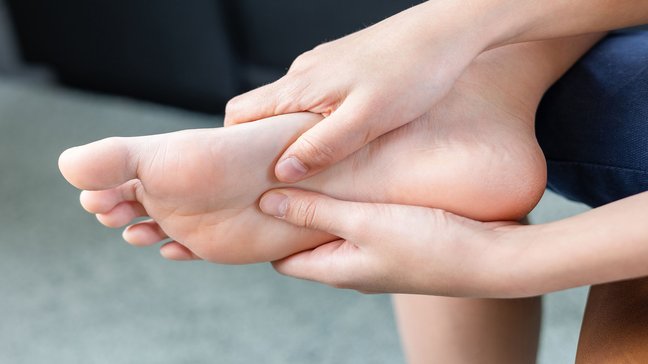
Metatarsalgia
Metatarsalgia describes pain in the forefoot area, often occurring under the metatarsal heads. These complaints usually arise from overuse, misalignments, or degenerative changes in the foot. People who stand a lot, walk, or are physically active are…
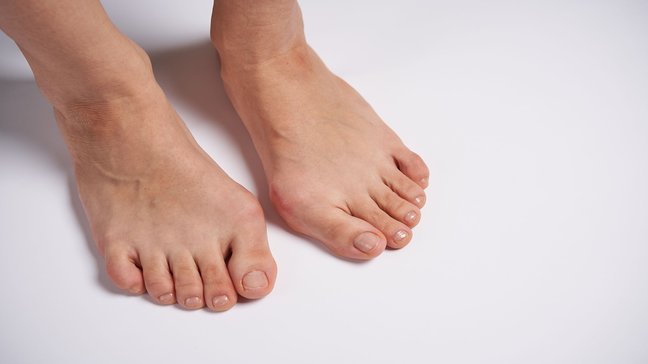
Hallux rigidus
Hallux rigidus is osteoarthritis of the big toe joint, which can lead to painful movement restrictions and eventually to joint stiffening. This condition significantly impairs the rolling motion of the foot and often affects people from middle age…
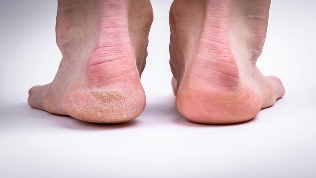
Haglund's heel (Haglund deformity)
The Haglund heel, also known as Haglund's exostosis or Haglund's syndrome, is a bony protrusion at the upper rear edge of the heel bone (calcaneus). This deformity often leads to pain and inflammation in the area of the Achilles tendon and the…

Fallen/Spread Foot
The flat/splayfoot is a common foot deformity characterized by a flattening of the longitudinal arch (flatfoot) and/or a widening of the forefoot due to a lowered transverse arch (splayfoot). These deformities often occur together and can lead to…
Clubfoot (Pes Equinovarus)
Classification of Clubfoot
Clubfoot is divided into different categories, depending on the cause and severity:
- Idiopathic clubfoot:
- Most common form with no known cause.
- Syndromic clubfoot:
- Occurs in connection with genetic syndromes such as arthrogryposis or trisomy 18.
- Neuropathic clubfoot:
- Caused by neurological disorders such as spina bifida.
- Postural clubfoot:
- Result of fetal position anomalies in the womb.
Causes of clubfoot
The exact causes of clubfoot are not fully understood. However, various factors play a role:
- Genetic predisposition:
- Clubfoot occurs more frequently in families, indicating a genetic component.
- Mechanical factors:
- Lack of space in the womb or an unfavorable position of the fetus can affect the development of the foot.
- Neurological causes:
- Conditions such as spina bifida or muscular dystrophy can affect foot positioning.
- Syndromic associations:
- Clubfoot can be part of a genetic syndrome.
Symptoms of clubfoot
The typical symptoms of a clubfoot are recognizable at birth:
- **Equinus position (Pes equinus):**
- The heel is elevated, and the foot cannot be placed flat.
- **Metatarsus adductus (Pes adductus):**
- The forefoot is turned inward.
- **Cavus foot (Pes cavus):**
- Excessive arching of the foot arch.
- **Varus position:**
- The foot is tilted inward, so the sole faces medially.
- **Limited mobility:**
- The foot cannot be actively brought into a normal position.
Diagnosis of clubfoot
The diagnosis is usually made immediately after birth through clinical examination. In some cases, clubfoot can already be detected prenatally using ultrasound. Further diagnostic measures include:
- Clinical assessment:
- Evaluation of the malposition and mobility of the foot.
- Imaging techniques:
- X-rays: Used for precise assessment of bone structure and joint position.
- Ultrasound: Non-invasive method for examining soft tissues and joints.
Treatment options for clubfoot
Conservative Therapy
The conservative treatment usually begins immediately after birth:
- Ponseti Method:
- A gradual manual correction of the foot, followed by plaster casts that are changed weekly. After about 5–8 weeks, a percutaneous Achilles tendon tenotomy is often performed to correct the remaining equinus position. The follow-up treatment involves special braces to prevent relapses.
- French Method:
- This method combines daily mobilization, bandages, and casts. It requires intensive cooperation from the parents.
Surgical Therapy
If conservative therapy is not sufficient, surgery may be necessary:
- Tendon Lengthening:
- Shortened tendons are surgically lengthened to improve the mobility of the foot.
- Osteotomies:
- Correction of misalignments of the foot bones through surgical procedures.
Preventive measures for clubfoot
Since the exact causes are not fully known, there are no specific preventive measures. However, it is important:
- Early diagnosis: Prenatal examinations can provide indications of clubfoot formation.
- Prompt treatment: An early start of therapy significantly increases the chances of success.
- Consistent follow-up treatment: Regular check-ups and wearing braces or orthoses reduce the risk of relapse.
Metatarsalgia
Metatarsalgia describes pain in the forefoot area, often occurring under the metatarsal heads. These complaints usually arise from overuse, misalignments, or degenerative changes in the foot. People who stand a lot, walk, or are physically active are…
Hallux rigidus
Hallux rigidus is osteoarthritis of the big toe joint, which can lead to painful movement restrictions and eventually to joint stiffening. This condition significantly impairs the rolling motion of the foot and often affects people from middle age…
Haglund's heel (Haglund deformity)
The Haglund heel, also known as Haglund's exostosis or Haglund's syndrome, is a bony protrusion at the upper rear edge of the heel bone (calcaneus). This deformity often leads to pain and inflammation in the area of the Achilles tendon and the…
Fallen/Spread Foot
The flat/splayfoot is a common foot deformity characterized by a flattening of the longitudinal arch (flatfoot) and/or a widening of the forefoot due to a lowered transverse arch (splayfoot). These deformities often occur together and can lead to…