

Calcaneal apophysitis
Classification of calcaneal apophysitis
Apophysitis calcanei can be divided into different degrees of severity:
- Mild course: Pain occurs only occasionally and with heavy exertion, and mobility remains largely preserved.
- Moderate course: Pain occurs even with moderate exertion, mobility is slightly restricted, and early signs of inflammation may be visible.
- Severe course: Severe pain even at rest, significant movement restrictions, and visible signs of inflammation such as swelling or redness are typical.
Causes of Apophysitis Calcanei
The main causes of calcaneal apophysitis can be divided into several categories:
- Mechanical overload:
- Repeated loads such as jumping, running, or sudden deceleration lead to microtrauma in the growth plate.
- Intense sports such as soccer, basketball, or track and field significantly increase the risk.
- Growth phases:
- During growth, there is often an asynchrony between bone growth and adaptation of the tendon-muscle unit. This makes the growth plate more susceptible to injuries and inflammation.
- Inappropriate footwear:
- Shoes without sufficient cushioning or stabilization can increase the load on the heel and worsen symptoms.
- Overweight:
- Increased mechanical load due to overweight increases the pressure on the growth plate and prolongs healing time.
Symptoms of Apophysitis Calcanei
Typical symptoms include:
- Heel pain: Especially during physical activity such as running or jumping, occasionally also at rest.
- Pressure sensitivity: Pain when touching or applying pressure to the heel.
- Limping: Children tend to limp to relieve the affected heel.
- Swelling and redness: In advanced cases, mild swelling or redness may occur, accompanied by local warmth.
- Movement restrictions: Difficulty bearing weight on the heel normally or performing movements such as rolling the foot.
Diagnosis of calcaneal apophysitis
Diagnosis is made through a thorough clinical examination and, if necessary, imaging:
- Medical history: The doctor obtains the medical history, focusing on sports activities, pain progression, and loading patterns.
- Physical examination: Palpation of the heel for tenderness and assessment of range of motion. Typical pain points along the growth plate are often identified.
- Imaging:
- X-ray: Visualization of the growth plate to rule out fractures or other bone disorders.
- MRI: Detailed analysis if more complex damage or inflammation is suspected in adjacent structures.
Treatment options for calcaneal apophysitis
Conservative Therapy
Most cases can be treated without surgical interventions:
- Immobilization:
- Reduction or pausing of sports activities to relieve the growth plate and promote healing.
- Heel pads and insoles:
- Special insoles or pads reduce pressure on the heel and reduce pain.
- Physiotherapy:
- Stretching exercises: Regular stretching of the calf muscles and Achilles tendon to reduce the tensile forces on the heel.
- Strengthening exercises: Promoting foot stability through targeted strengthening of the surrounding muscles.
- Pain therapy:
- Use of anti-inflammatory medications or cooling ointments to relieve acute symptoms.
- Cooling:
- Local cooling of the affected region to reduce inflammation and pain.
Surgical Therapy
Surgical treatment is extremely rare and is only considered in cases of persistent symptoms:
- Surgical intervention: Removal of inflamed tissue or correction of structural anomalies. These measures are used only in cases of complications or chronic cases.
Preventive measures for Apophysitis Calcanei
To prevent calcaneal apophysitis, the following measures are recommended:
- Appropriate footwear: Shoes with sufficient cushioning and good foot support reduce the load on the growth plate.
- Load management: Regular breaks and recovery periods during sports activities prevent overuse.
- Warm-up and stretching: Preparing the muscles through targeted warm-up and stretching before sports.
- Body weight control: Avoiding excess weight to reduce mechanical stress.
- Regular foot examinations: Early detection of symptoms through regular check-ups with a pediatrician or orthopedist.
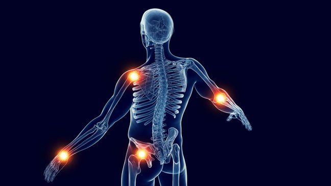
Osteonecrosis in childhood
Osteonecroses in childhood are conditions in which there is a localized blood flow disorder in the bone, leading to the death of the affected tissue. These disorders often occur at the growth zones (epiphyses) and are typically associated with pain,…

Transient synovitis
Transient synovitis of the hip, medically referred to as Coxitis fugax, is a temporary, non-bacterial inflammation of the hip joint. This condition primarily affects children between the ages of three and ten, with boys being more frequently affected…
Epiphysiolysis of the femoral head
Epiphysiolysis capitis femoris (ECF), also referred to as juvenile femoral head slippage, is an orthopedic condition in which the femoral head slips along the growth plate (epiphyseal plate) of the femur. This condition typically occurs in…
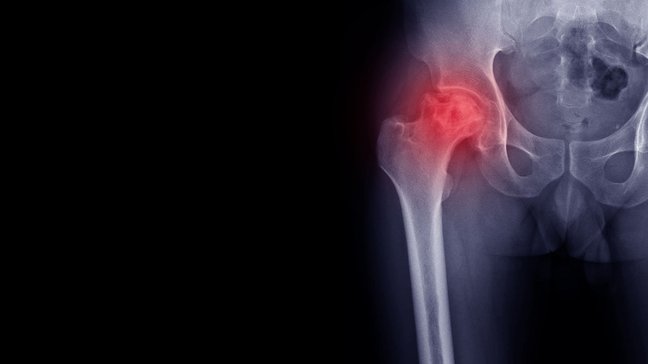
Perthes disease
Morbus Perthes, also known as Perthes disease, is a rare disorder of the hip joint in children. It arises from a temporary interruption of the blood supply to the femoral head, which leads to the death of bone tissue (aseptic necrosis of the femoral…
Calcaneal apophysitis
Classification of calcaneal apophysitis
Apophysitis calcanei can be divided into different degrees of severity:
- Mild course: Pain occurs only occasionally and with heavy exertion, and mobility remains largely preserved.
- Moderate course: Pain occurs even with moderate exertion, mobility is slightly restricted, and early signs of inflammation may be visible.
- Severe course: Severe pain even at rest, significant movement restrictions, and visible signs of inflammation such as swelling or redness are typical.
Causes of Apophysitis Calcanei
The main causes of calcaneal apophysitis can be divided into several categories:
- Mechanical overload:
- Repeated loads such as jumping, running, or sudden deceleration lead to microtrauma in the growth plate.
- Intense sports such as soccer, basketball, or track and field significantly increase the risk.
- Growth phases:
- During growth, there is often an asynchrony between bone growth and adaptation of the tendon-muscle unit. This makes the growth plate more susceptible to injuries and inflammation.
- Inappropriate footwear:
- Shoes without sufficient cushioning or stabilization can increase the load on the heel and worsen symptoms.
- Overweight:
- Increased mechanical load due to overweight increases the pressure on the growth plate and prolongs healing time.
Symptoms of Apophysitis Calcanei
Typical symptoms include:
- Heel pain: Especially during physical activity such as running or jumping, occasionally also at rest.
- Pressure sensitivity: Pain when touching or applying pressure to the heel.
- Limping: Children tend to limp to relieve the affected heel.
- Swelling and redness: In advanced cases, mild swelling or redness may occur, accompanied by local warmth.
- Movement restrictions: Difficulty bearing weight on the heel normally or performing movements such as rolling the foot.
Diagnosis of calcaneal apophysitis
Diagnosis is made through a thorough clinical examination and, if necessary, imaging:
- Medical history: The doctor obtains the medical history, focusing on sports activities, pain progression, and loading patterns.
- Physical examination: Palpation of the heel for tenderness and assessment of range of motion. Typical pain points along the growth plate are often identified.
- Imaging:
- X-ray: Visualization of the growth plate to rule out fractures or other bone disorders.
- MRI: Detailed analysis if more complex damage or inflammation is suspected in adjacent structures.
Treatment options for calcaneal apophysitis
Conservative Therapy
Most cases can be treated without surgical interventions:
- Immobilization:
- Reduction or pausing of sports activities to relieve the growth plate and promote healing.
- Heel pads and insoles:
- Special insoles or pads reduce pressure on the heel and reduce pain.
- Physiotherapy:
- Stretching exercises: Regular stretching of the calf muscles and Achilles tendon to reduce the tensile forces on the heel.
- Strengthening exercises: Promoting foot stability through targeted strengthening of the surrounding muscles.
- Pain therapy:
- Use of anti-inflammatory medications or cooling ointments to relieve acute symptoms.
- Cooling:
- Local cooling of the affected region to reduce inflammation and pain.
Surgical Therapy
Surgical treatment is extremely rare and is only considered in cases of persistent symptoms:
- Surgical intervention: Removal of inflamed tissue or correction of structural anomalies. These measures are used only in cases of complications or chronic cases.
Preventive measures for Apophysitis Calcanei
To prevent calcaneal apophysitis, the following measures are recommended:
- Appropriate footwear: Shoes with sufficient cushioning and good foot support reduce the load on the growth plate.
- Load management: Regular breaks and recovery periods during sports activities prevent overuse.
- Warm-up and stretching: Preparing the muscles through targeted warm-up and stretching before sports.
- Body weight control: Avoiding excess weight to reduce mechanical stress.
- Regular foot examinations: Early detection of symptoms through regular check-ups with a pediatrician or orthopedist.
Osteonecrosis in childhood
Osteonecroses in childhood are conditions in which there is a localized blood flow disorder in the bone, leading to the death of the affected tissue. These disorders often occur at the growth zones (epiphyses) and are typically associated with pain,…
Transient synovitis
Transient synovitis of the hip, medically referred to as Coxitis fugax, is a temporary, non-bacterial inflammation of the hip joint. This condition primarily affects children between the ages of three and ten, with boys being more frequently affected…
Epiphysiolysis of the femoral head
Epiphysiolysis capitis femoris (ECF), also referred to as juvenile femoral head slippage, is an orthopedic condition in which the femoral head slips along the growth plate (epiphyseal plate) of the femur. This condition typically occurs in…
Perthes disease
Morbus Perthes, also known as Perthes disease, is a rare disorder of the hip joint in children. It arises from a temporary interruption of the blood supply to the femoral head, which leads to the death of bone tissue (aseptic necrosis of the femoral…