

Infant hip ultrasound
Why is the examination important?
The development of the hip is not yet fully complete at birth. In about 4% of newborns, there is a maturation disorder of the hip.
Possible abnormalities include, for example:
• a hip dysplasia – the hip socket is not yet sufficiently developed
• a hip dislocation – the femoral head is not correctly positioned in the hip socket
• a altered angular position of the hip joint
If such a change is detected early, treatment can usually be very simple.
Hip ultrasound in the Mother-Child Pass
In Austria, hip ultrasound is an integral part of preventive examinations.
The Mother-Child Passport provides for:
• a first examination in the first week of life
• a follow-up examination at the age of 6 to 8 weeks
These examinations serve to assess the development of the hip joint at an early stage.
Advantages of the ultrasound examination
Hip ultrasound is especially suitable for infants.
• painless examination
• no radiation exposure
• can be repeated as often as needed for follow-up monitoring
• early assessment of hip development
With the help of ultrasound, the shape and maturity of the hip joint can be accurately assessed.
Detected early – well treatable
If hip dysplasia is diagnosed early, the development of the hip joint can be specifically supported.
• treatment begins early
• the duration of treatment is usually shorter
• surgical procedures can often be avoided
In most cases, consistent treatment leads to complete healing.
Important: Early diagnosis
The most important prerequisite for successful treatment is an early and accurate diagnosis. Hip ultrasound enables a reliable assessment of hip development already in the first weeks of life.
No. The examination is completely painless and is not stressful for your baby. Ultrasound does not use radiation and is therefore particularly well suited for infants.
In the Mother-Child Pass, two examinations are scheduled:
• a first examination in the first week of life
• a check-up at the age of 6 to 8 weeks
These appointments help to assess the development of the hip at an early stage.
A newborn’s hip is not yet fully developed at birth. Hip ultrasound can detect possible developmental abnormalities early, so that treatment can begin in a timely manner.
If hip dysplasia is detected early, the development of the hip joint can be specifically supported. In many cases, a simple treatment is sufficient to allow the hip to mature fully.
The examination usually only takes a few minutes. Your baby lies comfortably on an examination table while the hip joint is examined using ultrasound.

Orthopedic Body Check
Our orthopedic Body Check is a comprehensive examination to assess the entire musculoskeletal system. The goal is to identify improper loading, muscular imbalances, and potential injury risks at an early stage.
The Body Check is particularly…

Developmental monitoring in childhood
Orthopedic developmental monitoring helps to regularly check the growth and physical development of children. The goal is to detect possible postural problems or abnormalities of the musculoskeletal system at an early stage and, if necessary, to…
Hip dysplasia
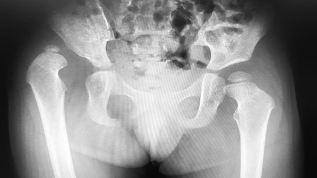
Hip dysplasia, also referred to as hip joint dysplasia, is a congenital or acquired malformation of the hip joint. The hip socket (acetabulum) is insufficiently developed, which leads to the femoral head (caput femoris) not sitting optimally in the…
Infant hip ultrasound
Why is the examination important?
The development of the hip is not yet fully complete at birth. In about 4% of newborns, there is a maturation disorder of the hip.
Possible abnormalities include, for example:
• a hip dysplasia – the hip socket is not yet sufficiently developed
• a hip dislocation – the femoral head is not correctly positioned in the hip socket
• a altered angular position of the hip joint
If such a change is detected early, treatment can usually be very simple.
Hip ultrasound in the Mother-Child Pass
In Austria, hip ultrasound is an integral part of preventive examinations.
The Mother-Child Passport provides for:
• a first examination in the first week of life
• a follow-up examination at the age of 6 to 8 weeks
These examinations serve to assess the development of the hip joint at an early stage.
Advantages of the ultrasound examination
Hip ultrasound is especially suitable for infants.
• painless examination
• no radiation exposure
• can be repeated as often as needed for follow-up monitoring
• early assessment of hip development
With the help of ultrasound, the shape and maturity of the hip joint can be accurately assessed.
Detected early – well treatable
If hip dysplasia is diagnosed early, the development of the hip joint can be specifically supported.
• treatment begins early
• the duration of treatment is usually shorter
• surgical procedures can often be avoided
In most cases, consistent treatment leads to complete healing.
Important: Early diagnosis
The most important prerequisite for successful treatment is an early and accurate diagnosis. Hip ultrasound enables a reliable assessment of hip development already in the first weeks of life.
No. The examination is completely painless and is not stressful for your baby. Ultrasound does not use radiation and is therefore particularly well suited for infants.
In the Mother-Child Pass, two examinations are scheduled:
• a first examination in the first week of life
• a check-up at the age of 6 to 8 weeks
These appointments help to assess the development of the hip at an early stage.
A newborn’s hip is not yet fully developed at birth. Hip ultrasound can detect possible developmental abnormalities early, so that treatment can begin in a timely manner.
If hip dysplasia is detected early, the development of the hip joint can be specifically supported. In many cases, a simple treatment is sufficient to allow the hip to mature fully.
The examination usually only takes a few minutes. Your baby lies comfortably on an examination table while the hip joint is examined using ultrasound.
Orthopedic Body Check
Our orthopedic Body Check is a comprehensive examination to assess the entire musculoskeletal system. The goal is to identify improper loading, muscular imbalances, and potential injury risks at an early stage.
The Body Check is particularly…
Developmental monitoring in childhood
Orthopedic developmental monitoring helps to regularly check the growth and physical development of children. The goal is to detect possible postural problems or abnormalities of the musculoskeletal system at an early stage and, if necessary, to…
Hip dysplasia
Hip dysplasia, also referred to as hip joint dysplasia, is a congenital or acquired malformation of the hip joint. The hip socket (acetabulum) is insufficiently developed, which leads to the femoral head (caput femoris) not sitting optimally in the…