

Avascular necrosis of the femoral head
Classification of Femoral Head Necrosis
Hip head necrosis is classified according to the cause and progression of the disease:
- Traumatic hip head necrosis:
- Caused by injuries such as femoral neck fractures or hip dislocations.
- Non-traumatic hip head necrosis:
- Occurs without prior trauma, often due to systemic diseases or medication intake.
- Early stage:
- The bone shows no visible changes yet, symptoms are often nonspecific.
- Advanced stage:
- Bone decay and collapse of the hip head with significant restriction of joint function.
Causes of femoral head necrosis
The causes of femoral head necrosis are diverse and include:
- Traumatic events:
- Injuries such as femoral neck fractures or hip dislocations that interrupt the blood supply to the femoral head.
- Systemic diseases:
- Diseases such as diabetes mellitus or systemic lupus erythematosus impair the microcirculation in the bone.
- Medications:
- Long-term use of high-dose corticosteroids is a known risk factor.
- Alcohol consumption:
- Excessive alcohol consumption damages blood vessels and increases the risk of necrosis.
- Blood clotting disorders:
- Conditions such as sickle cell anemia or thrombosis can impair the blood flow to the femoral head.
Symptoms of femoral head necrosis
The symptoms of femoral head necrosis depend on the stage of the disease and can begin insidiously:
- Pain:
- Groin pain that can radiate to the thigh or buttocks.
- Pain often occurs first during exertion and later also at rest.
- Movement restrictions:
- Difficulty walking, climbing stairs, or bending over.
- Loss of strength:
- Weakness in the affected leg due to protective posture and muscle atrophy.
- Changes in gait:
- Affected individuals often limp to avoid pain.
- Grinding or rubbing:
- Mechanical noises in the hip joint during movement in the advanced stage.
Diagnosis of femoral head necrosis
The diagnosis of femoral head necrosis is based on medical history, clinical examination, and imaging techniques:
- Medical history:
- The doctor inquires about possible risk factors such as alcohol or medication consumption and previous injuries.
- Clinical examination:
- Checking for pain points, mobility, and gait pattern.
- Imaging techniques:
- X-ray: Depiction of bone changes and joint space narrowing in the advanced stage.
- MRI: Early detection of circulatory disorders and necrosis areas in the bone.
- Bone scintigraphy: Depiction of metabolic activities in the bone in case of unclear diagnosis.
Treatment options for femoral head necrosis
Conservative Therapy
In early stages, conservative treatment can alleviate symptoms and slow the progression of the disease:
- Physiotherapy:
- Exercises for mobilization and strengthening of the muscles.
- Pain Therapy:
- Use of non-steroidal anti-inflammatory drugs (NSAIDs) for pain relief.
- Joint Relief:
- Use of walking aids or relief orthoses to protect the hip joint.
- Shock Wave Therapy:
- Promotion of blood circulation and regeneration of the bone.
Surgical Therapy
In advanced femoral head necrosis, surgery is often unavoidable:
- Decompression:
- Surgical relief of the bone interior to improve blood circulation.
- Bone Transplantation:
- Replacement of necrotic tissue with healthy bone material.
- Hip Endoprosthesis:
- Replacement of the hip joint with a prosthesis in case of irreparable damage.
Preventive measures for femoral head necrosis
To prevent femoral head necrosis, the following measures are recommended:
- Avoidance of risk factors: Limiting alcohol consumption and avoiding unnecessary medication intake.
- Regular exercise: Promoting circulation through joint-friendly sports such as swimming or cycling.
- Weight management: Reducing excess weight to relieve the hip joint.
- Early treatment: Immediate clarification of hip pain or risk factors by a doctor.
- Education: Knowledge about possible triggers and timely action at the first symptoms.
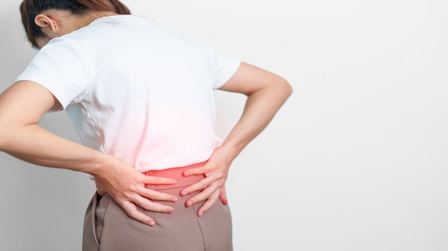
Iliopsoas shortening
The iliopsoas shortening describes a shortening of the iliopsoas muscle, which consists of the psoas major muscle and the iliacus muscle. This muscle complex is the main flexor of the hip joint and plays an important role in stabilizing the spine. A…
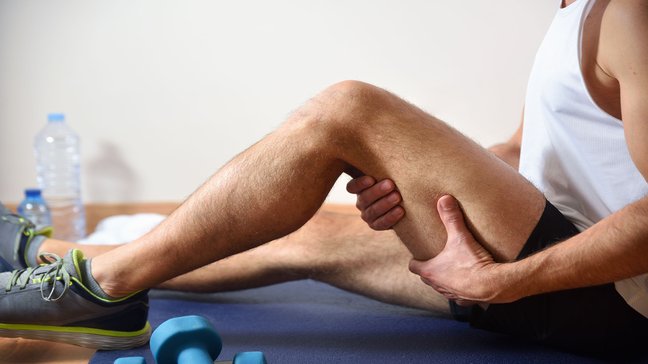
Adductor strain
An adductor strain is a common sports injury caused by overstretching or overloading the adductor muscles on the inner thigh. This muscle group is responsible for bringing the leg toward the body's midline and plays a crucial role in movements such…
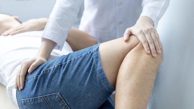
Labral tear of the hip
A labral tear of the hip refers to damage or injury to the acetabular labrum, a cartilage-like structure that surrounds the hip socket. The labrum stabilizes the hip joint by increasing the contact area between the socket and the head while…
Avascular necrosis of the femoral head
Classification of Femoral Head Necrosis
Hip head necrosis is classified according to the cause and progression of the disease:
- Traumatic hip head necrosis:
- Caused by injuries such as femoral neck fractures or hip dislocations.
- Non-traumatic hip head necrosis:
- Occurs without prior trauma, often due to systemic diseases or medication intake.
- Early stage:
- The bone shows no visible changes yet, symptoms are often nonspecific.
- Advanced stage:
- Bone decay and collapse of the hip head with significant restriction of joint function.
Causes of femoral head necrosis
The causes of femoral head necrosis are diverse and include:
- Traumatic events:
- Injuries such as femoral neck fractures or hip dislocations that interrupt the blood supply to the femoral head.
- Systemic diseases:
- Diseases such as diabetes mellitus or systemic lupus erythematosus impair the microcirculation in the bone.
- Medications:
- Long-term use of high-dose corticosteroids is a known risk factor.
- Alcohol consumption:
- Excessive alcohol consumption damages blood vessels and increases the risk of necrosis.
- Blood clotting disorders:
- Conditions such as sickle cell anemia or thrombosis can impair the blood flow to the femoral head.
Symptoms of femoral head necrosis
The symptoms of femoral head necrosis depend on the stage of the disease and can begin insidiously:
- Pain:
- Groin pain that can radiate to the thigh or buttocks.
- Pain often occurs first during exertion and later also at rest.
- Movement restrictions:
- Difficulty walking, climbing stairs, or bending over.
- Loss of strength:
- Weakness in the affected leg due to protective posture and muscle atrophy.
- Changes in gait:
- Affected individuals often limp to avoid pain.
- Grinding or rubbing:
- Mechanical noises in the hip joint during movement in the advanced stage.
Diagnosis of femoral head necrosis
The diagnosis of femoral head necrosis is based on medical history, clinical examination, and imaging techniques:
- Medical history:
- The doctor inquires about possible risk factors such as alcohol or medication consumption and previous injuries.
- Clinical examination:
- Checking for pain points, mobility, and gait pattern.
- Imaging techniques:
- X-ray: Depiction of bone changes and joint space narrowing in the advanced stage.
- MRI: Early detection of circulatory disorders and necrosis areas in the bone.
- Bone scintigraphy: Depiction of metabolic activities in the bone in case of unclear diagnosis.
Treatment options for femoral head necrosis
Conservative Therapy
In early stages, conservative treatment can alleviate symptoms and slow the progression of the disease:
- Physiotherapy:
- Exercises for mobilization and strengthening of the muscles.
- Pain Therapy:
- Use of non-steroidal anti-inflammatory drugs (NSAIDs) for pain relief.
- Joint Relief:
- Use of walking aids or relief orthoses to protect the hip joint.
- Shock Wave Therapy:
- Promotion of blood circulation and regeneration of the bone.
Surgical Therapy
In advanced femoral head necrosis, surgery is often unavoidable:
- Decompression:
- Surgical relief of the bone interior to improve blood circulation.
- Bone Transplantation:
- Replacement of necrotic tissue with healthy bone material.
- Hip Endoprosthesis:
- Replacement of the hip joint with a prosthesis in case of irreparable damage.
Preventive measures for femoral head necrosis
To prevent femoral head necrosis, the following measures are recommended:
- Avoidance of risk factors: Limiting alcohol consumption and avoiding unnecessary medication intake.
- Regular exercise: Promoting circulation through joint-friendly sports such as swimming or cycling.
- Weight management: Reducing excess weight to relieve the hip joint.
- Early treatment: Immediate clarification of hip pain or risk factors by a doctor.
- Education: Knowledge about possible triggers and timely action at the first symptoms.
Iliopsoas shortening
The iliopsoas shortening describes a shortening of the iliopsoas muscle, which consists of the psoas major muscle and the iliacus muscle. This muscle complex is the main flexor of the hip joint and plays an important role in stabilizing the spine. A…
Adductor strain
An adductor strain is a common sports injury caused by overstretching or overloading the adductor muscles on the inner thigh. This muscle group is responsible for bringing the leg toward the body's midline and plays a crucial role in movements such…
Labral tear of the hip
A labral tear of the hip refers to damage or injury to the acetabular labrum, a cartilage-like structure that surrounds the hip socket. The labrum stabilizes the hip joint by increasing the contact area between the socket and the head while…